Neurodegenerative disease

A blood test to diagnose ALS.

A blood test to diagnose amyotrophic lateral sclerosis (ALS) could be widely available within two years.

Currently, ALS is diagnosed through a clinical examination by a neurologist but distinguishing it from other neurological diseases requires tracking symptom progression.

 This is problematic, as the average survival time in ALS is approximately three years, meaning many patients deteriorate significantly before receiving a definitive diagnosis.

Initial misdiagnosis rates range as high as 68%, delaying treatment and causing patients to be passed between specialists, increasing anxiety, unnecessary interventions, and costs.

A simple blood test for ALS would be a game-changer. It would speed up diagnosis, reduce anxiety, lower costs, and support the development of new drugs. Given the high misdiagnosis rate, a negative result would also be highly valuable.

Extracellular vesicles are nano-sized particles that circulate in the blood, are secreted by all cells, an contain microRNA. 

Researchers at the Brain Chemistry Labs in Jackson, Wyoming, reported today in Brain Communications that they have identified an ALS-specific biomarker, an “ALS fingerprint,” in the blood. The biomarker of eight microRNAs (miRNAs) can be detected via a simple blood draw.

Using next-generation sequencing and real-time PCR, the team analyzed blood samples from patients with ALS, Primary Lateral Sclerosis (PLS), Parkinson’s disease (PD), and healthy individuals.

The eight microRNA ALS fingerprint accurately detects ALS with as high as 98% accuracy and can separate ALS from PLS and PD.

“Faster diagnoses will allow for earlier treatment, which will improve patient outcomes,” says Sandra Banack, lead author of the study.

To confirm its reliability, the biomarker was tested across four different patient groups, in two labs, with various technicians and collection methods. The ALS fingerprint consistently produced reliable results.

Researchers believe this blood test could assist neurologists in diagnosing ALS and complement current clinical assessments.

Dr. Paul Alan Cox, Executive Director of Brain Chemistry Labs, hopes to secure a diagnostic company partnership and make this test widely available to neurologists within 18 to 24 months.

————

Brain Chemistry Labs is a 501(c)(3) not-for-profit organization in Jackson, Wyoming, that seeks to discover new ways to prevent, diagnose and treat ALS, Alzheimer’s, Parkinson’s and other serious brain diseases.

The Brain Communications paper, “A microRNA diagnostic biomarker for amyotrophic lateral sclerosis” (DOI 10.1093/braincomms/fcae268) can be accessed here.

Neurotoxin BMAA found in dust from Great Salt Lake.

In a startling discovery, researchers have identified a chronic neurotoxin known as BMAA in dust particles from the Great Salt Lake's dried lakebed. This toxin, linked to neurodegenerative illnesses, has become a significant health concern due to its presence in windblown dust that reaches populous metropolitan areas along the Wasatch Front.

Dr James Metcalf collects dust samples from the Great Salt Lake.

The dust, enriched with heavy metals and now with identified cyanobacteria and their toxins, poses an unsettling environmental health risk.

Studies have indicated that chronic dietary exposure to BMAA can trigger ALS-type neuropathology, with the neurotoxin now considered the most substantial environmental risk factor for developing Amyotrophic Lateral Sclerosis (ALS).

Although BMAA exposure also causes laboratory animals to form Alzheimer’s-type neuropathology, its role in Alzheimer’s, as well as Parkinson’s, is not fully understood.

However, its connection to ALS has been corroborated by two recent epidemiological studies, marking it as the most robust environmental link to the disease thus far.

Notably, Dartmouth researchers observed a higher risk of ALS in residents living near cyanobacteria-infested rivers and lakes in New Hampshire, underscoring the potential hazard of BMAA exposure.

Studies have indicated that chronic dietary exposure to BMAA can trigger ALS-type neuropathology, with the neurotoxin now considered the most substantial environmental risk factor for developing Amyotrophic Lateral Sclerosis (ALS).

Furthering the concern, the nonprofit Brain Chemistry Labs in Jackson Hole has been monitoring BMAA and other cyanotoxins in water bodies like Lake Okeechobee and collaborating with the Calusa Water Keeper to study airborne cyanobacterial toxins in Florida.

International research groups from Sweden, China, and France are also investigating chronic BMAA exposure as an ALS risk factor.

While ninety percent of ALS cases are sporadic and ten percent familial, the threat posed by the inhalation of BMAA-laced dust remains unclear.

What is clear, however, is the urgent need for comprehensive studies to determine the potential increase in ALS risk due to exposure to the Great Salt Lake dust.

The study was published this week in the journal Toxins.

Cyanotoxin Analysis of Air Samples from the Great Salt Lake. James S. Metcalf, Sandra Anne Banack and Paul Alan Cox. Toxins 2023, 15(11), 659; https://doi.org/10.3390/toxins15110659


New Alzheimer’s Biomarker May Facilitate Rapid Diagnosis

Blood test collection tube saying "Alzheimer's Disease"

Discovery of a unique ratio of metabolites from blood samples of early-stage Alzheimer’s patients promises to speed diagnosis of Alzheimer’s disease.

Although symptoms of advanced Alzheimer’s disease are well known, diagnosis of Alzheimer’s disease in its earliest stages requires careful cognitive testing by neurologists.

Discovery of a unique ratio of metabolites from blood samples of early-stage Alzheimer’s patients promises to speed diagnosis, allowing earlier treatments to be initiated.

“We were delighted to discover that the ratio of two molecules, 2-aminoethyl dihydrogen phosphate and taurine, allows us to reliably discriminate samples of early-stage Alzheimer’s patients from controls,” said Dr. Sandra Banack, lead author of the report in PLOS ONE and Senior Scientist at the Brain Chemistry Labs in Jackson Hole.

Dr. Sandra Banack works on the amino acid analyzer in the Brain Chemistry Labs, Jackson, Wyoming.

The blood samples were drawn from patients enrolled in an FDA-approved Phase II trial at Dartmouth Hitchcock Medical Center in New Hampshire and then shipped to the Brain Chemistry Labs for analysis. Current attempts to diagnose Alzheimer’s disease from blood samples depend on the presence of amyloid fragments, the molecules that cause brain tangles and plaques.

“At the Brain Chemistry Labs, we consider amyloid plaques to be a consequence rather than the cause of Alzheimer’s disease,” Dr. Paul Alan Cox, Executive Director of the Brain Chemistry Labs explains. “What is exciting about this new discovery is that it does not depend on amyloid and the assay can be performed on analytical equipment that is already present in most large hospitals.”

Their report, written with Alzheimer’s expert Dr. Aleksandra Stark, “A Possible Blood Plasma Biomarker for Early-stage Alzheimer’s Disease” is being published this week in PLOS ONE.


About the Brain Chemistry Labs: The Brain Chemistry Labs is a not-for-profit research institute based in Jackson Hole focused on improving outcomes for patients suffering from Alzheimer’s, ALS, and other neurodegenerative illnesses.

Contacts:
Dr. Sandra Banack, Tel: 307-734-1680, sandra@ethnomedicine.org
Dr. Paul Alan Cox, Tel: 801-375-6214, paul@ethnomedicine.org

Download our press release here

Like Origami Paper, Proteins Must Be Folded Correctly

In Origami, 2-D pieces of paper can be folded into 3-D masterpieces. 

A protein is a linear sequence of amino acids linked together through peptide bonds. Proteins differ in the sequence of the twenty different amino acids that our cells assemble, and in their length. However, to function properly, the 1-D protein has to be correctly folded into a 3-D structure.

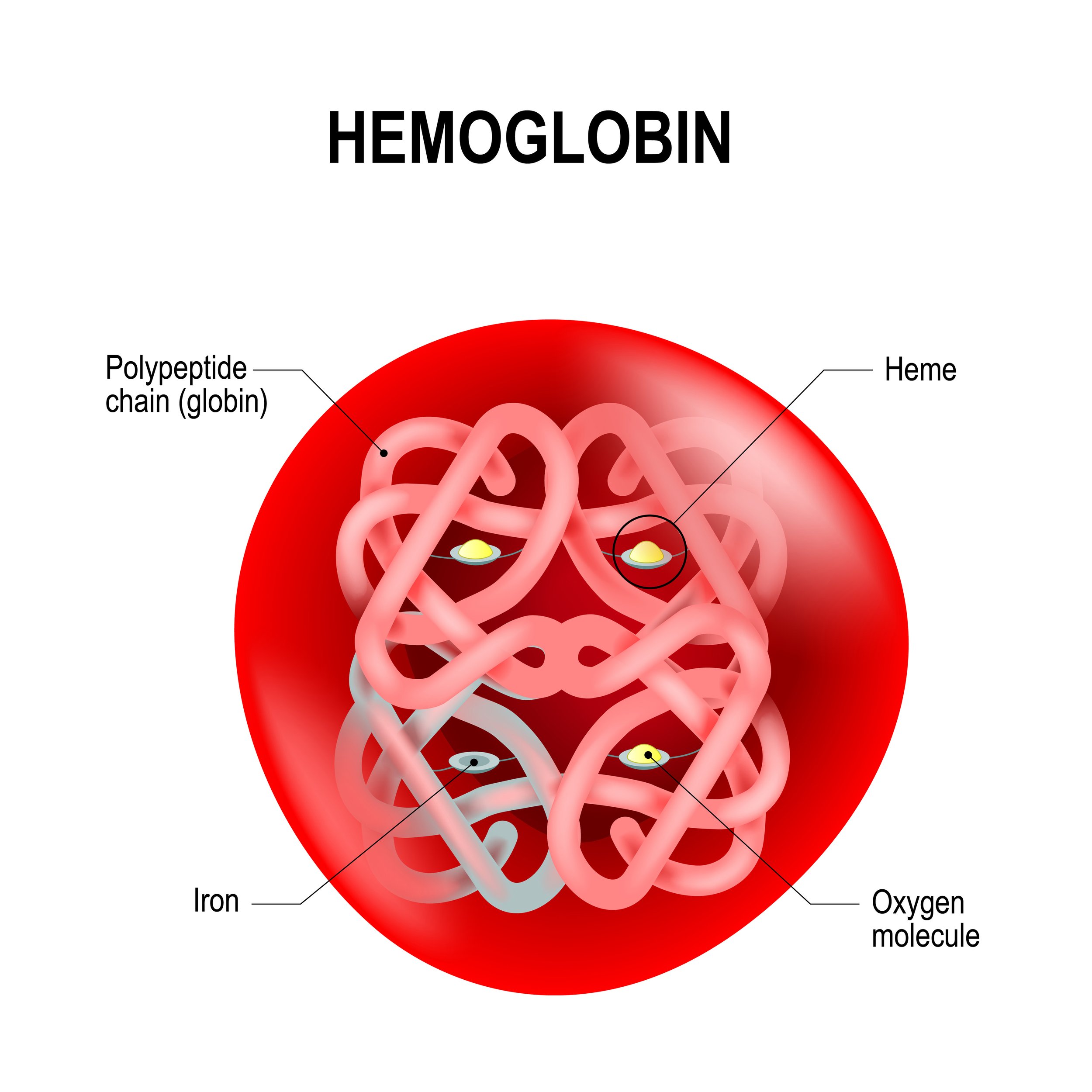
Hemoglobin (below) is the oxygen-carrying protein in our bloodstream, but it cannot carry oxygen unless its linear structure is folded properly. When a protein misfolds, it becomes toxic. Unlike other cells in the body, neurons cannot dilute the toxin by rapidly dividing. Misfolded proteins clog up transportation systems in neurons and cause them to die.

Each of the progressive neurodegenerative diseases is characterized by different proteins which misfold. Our strategy in developing new therapies for ALS, Alzheimer’s, and Parkinson’s is to find drugs that stop protein misfolding. We discovered that the amino acid L-serine helps to prevent protein misfolds.

Protein Misfolds Spread Like Prions in Alzheimer’s

The addition of L-serine to the diet of mice restored their memory

We believe protein misfolding to be the ultimate cause of Alzheimer’s, ALS, and other neurodegenerative diseases.

We discovered that environmental toxins such as BMAA can trigger protein misfolding.

How do protein misfolds spread from neuron to neuron? L-serine is produced within astrocytes, which help neurons.

Recently, a French team found that mice which cannot produce L-serine in astrocytes have memory problems. The addition of L-serine to their diet restored their memory.

Deficiencies in L-serine production within astrocytes may allow protein misfolds to propagate within the brain.

A New Approach to Parkinson’s Disease Therapy

David Long

Lewy bodies, formed from misfolded α-synuclein protein, are the neuropathological feature of Parkinson’s disease. While levodopa can help control symptoms, no current Parkinson’s medication slows disease progression.

 We attempted to produce Lewy bodies in vivo but did not have enough funds to continue the experiments. Enter Ty and Sue Measom of Logan, Utah, and Fred and Candy Berthrong of Providence, Utah who provided a generous gift to complete the study in which we found that BMAA triggers Lewy bodies in marmosets.

 We were unable to begin a second study until the philanthropic torch was picked up by Robert and Robin Paulson and Stan and Mary Seidler of Jackson Hole. They provided the lead gifts to determine if L-serine or L-tyrosine can slow Lewy body formation.

A generous pledge from David and Lisa Long of Lake Forest, Illinois allowed us to accelerate the project. Our colleagues at NeuroScience Associates in Tennessee assisted us by giving a significant discount on the regular fees for state-of-the-art sectioning and staining of the tissues. We still have $100,000 to raise to finish the project but are proceeding at full speed.

Because of the generous donors who helped us, including Bobbie Sweet, John Madigan, and James and Ellen Walton, we will have the final results in February 2022. If successful, this new drug will remain a lasting legacy for David Long who passed away on September 19, 2021.

Brain Chemistry Labs focus on Florida blooms.

Dr. James Metcalf samples the waters of Florida

Massive cyanobacterial blooms associated with the emergency release of nutrient-laden Lake Okeechobee water down the St. Lucie river in the summer of 2016 exposed Florida residents to a potent toxin called microcystin which is known to cause liver cancer. READ MORE.